
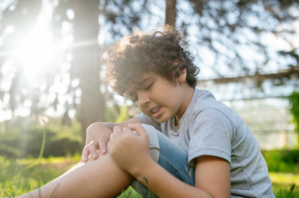
The knee is the largest joint in the human body, is extremely complex in structure, and is therefore prone to a wide range of diseases and injuries. Because children are taking up sport earlier and more intensively, the knees of children and adolescents are one of the more common reasons for a visit to a paediatric orthopaedic specialist. In sporting activities, the knee is most often the severely injured joint that requires surgical treatment. Likewise, children grow in height most “from the knee”, the growth plates there are the most active and cells divide fastest, which is why malignant bone diseases in children are, in fact, most common around the knee. However, benign conditions are far more common overall. Children are often told that pain around the knee is due to a growth spurt, and it is not unusual to hear parents in the clinic say that they, too, had similar knee problems as children and had to do exercises for it.
The knee joint consists of three bones: the lower end of the femur (thigh bone), the upper end of the tibia (shin bone), and the patella (kneecap). To improve the poor congruence – the fit – between the bones of the knee, crescent-shaped cartilage structures called menisci sit between the femur and tibia. Numerous ligaments in and around the knee, together with tendons and muscles, are responsible for giving the knee a full, stable range of motion. Each of these parts of the knee can be a source of trouble for a child, and therefore a reason to visit a paediatric orthopaedic specialist.
Examining the knee in children and adolescents
Every examination begins with a conversation with the child and their parent or adult companion to establish the reason for the visit. If pain is the issue, it is important to establish when it began, how long it has lasted, whether it worsens with activity, and whether there was any injury or particular moment when the knee pain started. Children often do not recall a traumatic event; instead, the pain develops gradually. The character of the pain also matters – severe, limiting pain differs from pain before falling asleep or only at night. It is important to ask about swelling or changes in skin colour around the knee, and whether, alongside pain, there is also limited movement in the knee, which could point to a meniscus injury. Particular attention should be paid to any feeling of knee instability, which can be a sign of an ACL tear or patellar instability.
The conversation is followed by inspection – observing the child's knee. The child should be in their underwear so the whole leg can be seen. The examiner looks at the alignment of the legs, whether there is a pronounced valgus (“knock-knee”) or varus (“bow-leg”) shape, or whether the knee is slightly bent so the child cannot fully straighten it. The contours of the knee, the position of the patella, and the presence of swelling, thickening or skin colour changes are noted. Wasting of the thigh muscles is a sign of prolonged knee weakness or pain and should be noted. Finally, the child's gait should be observed to check for a limp.
After the standing examination, the child lies down on the examination table, and palpation – feeling the individual parts of the knee – is used to locate areas of tenderness. It is sometimes useful to ask the child to point with a finger to the most painful spot. The patella is palpated and its stability checked, along with the ends of the femur and tibia, the joint line, and the back – popliteal – part of the knee. Tenderness at the tip of the patella points to Sinding-Larsen-Johansson disease, while tenderness over the tibial tuberosity (the bony bump below the front of the knee) points to Osgood-Schlatter disease. If a child has pain on palpation of the end of the femur in the knee, together with a feeling that something is catching in the joint, the cause may be osteochondritis dissecans. Palpation also establishes whether there is fluid in the knee, which can be a sign of bleeding following a meniscus injury, an ACL injury, or a patella dislocation. Fluid in the knee without a preceding injury may be a sign of transient synovitis, juvenile idiopathic arthritis, or septic arthritis.
The examination then moves on to specific tests of knee ligament stability, checking the condition of the anterior and posterior cruciate ligaments, the medial and lateral collateral ligaments, and the ligaments attached to the patella. The condition of the meniscus is checked with compression and rotation tests of the knee in various positions. The knee's range of motion in children runs from fully straight to around 150 degrees of bending. Some children have more pronounced ligament laxity and can extend the knee 5–10 degrees beyond straight. At this point, possible shortening of the thigh muscles, which can occur in children during phases of rapid growth, is also checked. Finally, every clinical knee examination should also include an assessment of hip range of motion, since children sometimes experience hip problems as pain around the knee.
After the clinical knee examination, if there is a need to clarify the problem or confirm particular suspicions, imaging of the knee is required. A knee ultrasound can be performed immediately in the clinic to confirm the presence of fluid, a popliteal cyst, or to assess the condition of the ligaments around the knee. The bones of the knee should be visualised by X-ray in at least two views – AP (anterior-posterior; front view) and LL (latero-lateral; side view) – and when patellar problems are suspected, an axial (skyline) view of the patella should also be taken. In cases of pronounced knock-knee or bow-leg deformity, standing panoramic X-rays of the lower limbs should be taken to assess the anatomical and mechanical leg axes. However, the knee consists of many ligamentous, tendinous and cartilaginous structures that are not visible on X-ray, so an MRI is needed for a proper assessment of their condition – for example, for meniscus, cartilage or cruciate ligament injuries.
Naturally, a visit to a paediatric orthopaedic specialist should always be considered from a medical standpoint. However, the social and psychological importance of the onset and development of a knee disorder in a child must not be overlooked. For example, a child with an unstable knee will not feel comfortable playing, will avoid company for fear of new injuries, and may ultimately withdraw and suffer in silence. For this reason too, it is important to act in time and bring the child in for an examination, in order to prevent further negative consequences from every possible perspective.
For more information or to schedule an appointment, please get in touch via the contact form.
Book Appointment